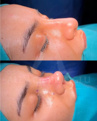
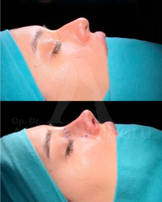
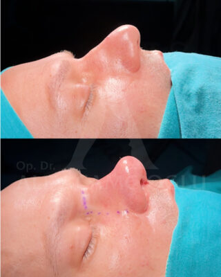
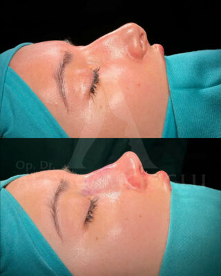
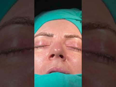
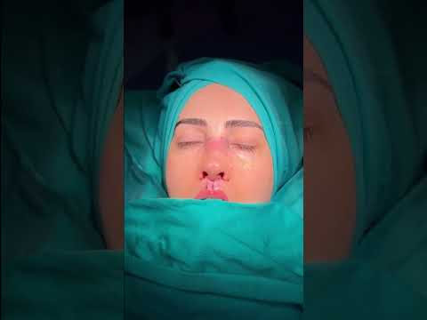
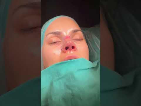
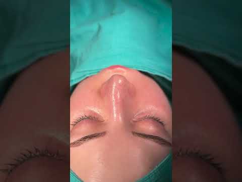
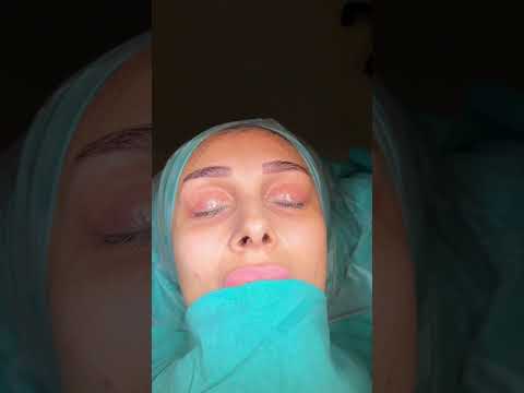
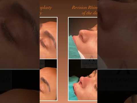
Functional Nose Surgery in Turkey
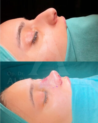
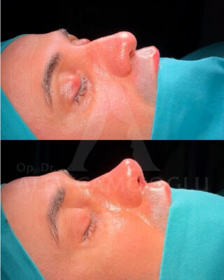
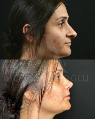
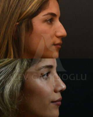
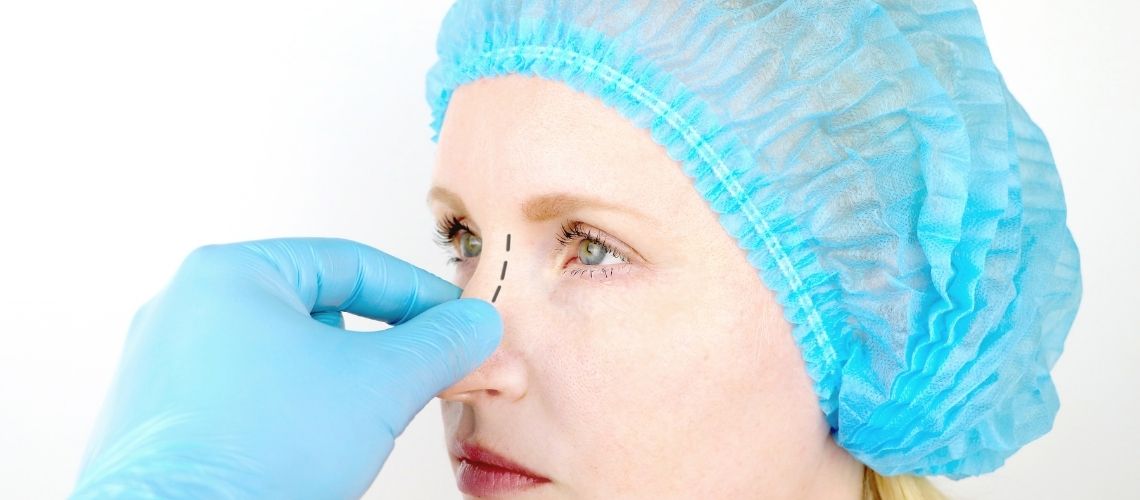
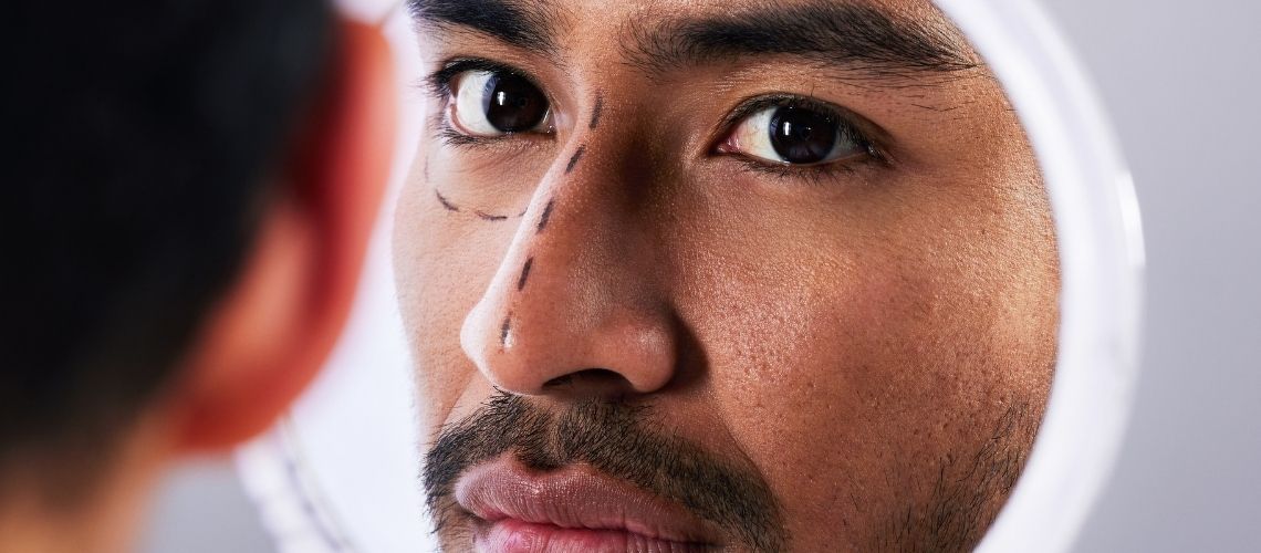
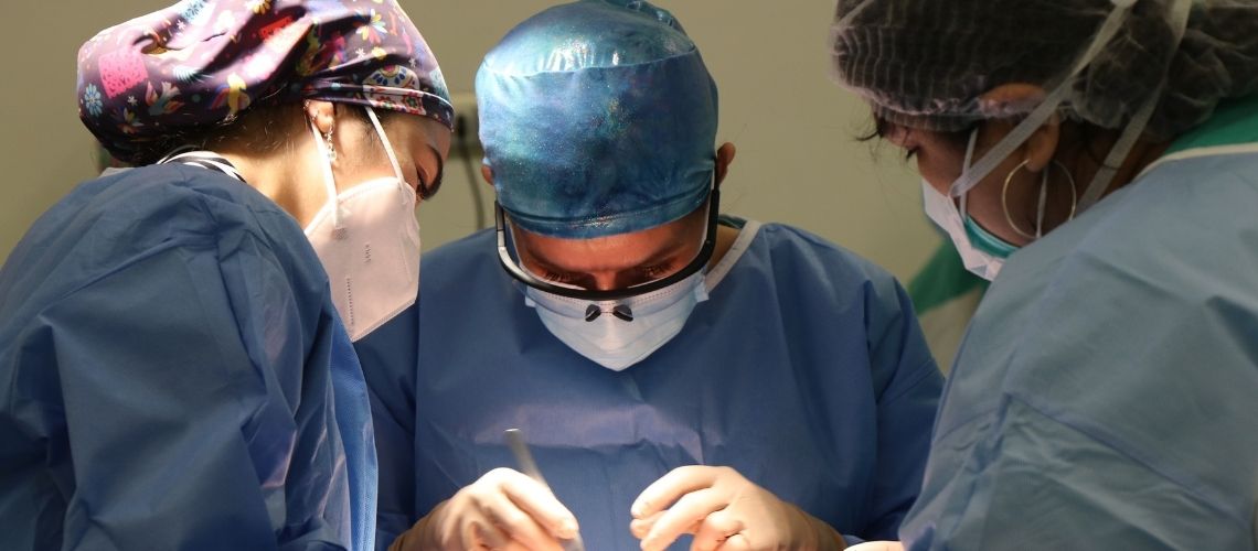
Improve both the appearance and function of your nose with Functional Rhinoplasty in Turkey at Dr. Camcıoğlu’s clinic. If you’re experiencing breathing difficulties or structural issues, we can address these concerns while also enhancing your nasal aesthetics. Our holistic approach ensures you enjoy both form and function improvements.
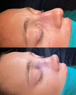
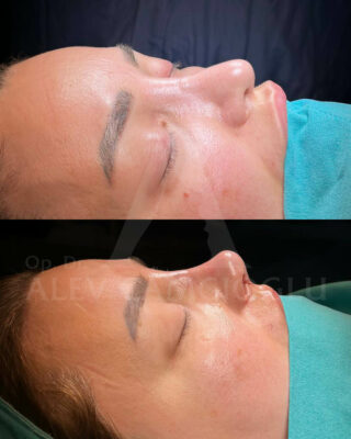
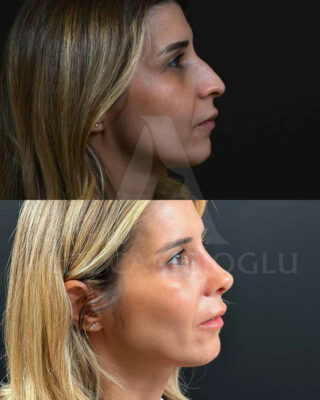
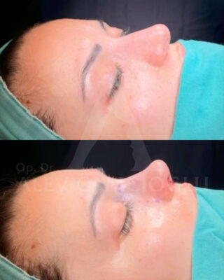
Cost of Functional Rhinoplasty in Istanbul, Turkey ranges between €5,000– €7,000 in 2025 depending on hospital, package, and anatomy of your nose. Check our prices, before & after photos, and patient results from the USA, UK, Canada, Australia, and the rest of the world.

Approaches to Functional Rhinoplasty
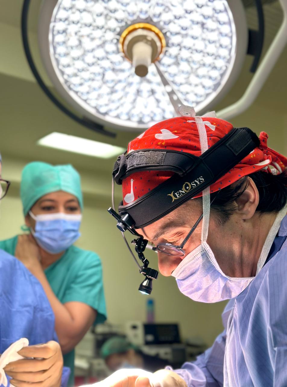
Functional rhinoplasty aims to improve the functionality of nasal structures. Chronic nasal congestion is a common issue that negatively affects the quality of life for many adults. Studies show that a significant part of this condition is due to nasal valve dysfunction. The nasal valve is considered the main area of resistance in nasal airflow. Nasal valve dysfunction is typically examined under two main categories: static and dynamic dysfunction.
Static dysfunction presents as congestion at rest, requiring more negative intranasal pressure for resolution. Dynamic dysfunction, on the other hand, results from insufficient structural support in the lateral nasal wall. This condition causes the valve to collapse at low transmural pressures.
Surgical techniques used to correct nasal valve dysfunction in both cases include:
- Spreader grafts: Widen the nasal valve area, relieving static congestion.
- Cartilaginous batten grafts: Used to prevent dynamic collapse of the lateral wall.
- Cosmetic rhinoplasties: Applied to patients with newly emerging symptoms.
Functional rhinoplasty is customized according to the specific needs of each patient. The choice of surgical methods varies depending on the patient’s anatomical structure and the nature of the existing problem. The procedure may be supported by medical treatment to address physiological issues such as mucosal inflammation.
Once mucosal swelling is resolved, structural defects in the nasal valve can be clearly assessed. In this case, surgical repair can be considered to maximize functional improvement. However, the patient’s condition should be carefully examined before surgery. This allows for distinguishing between static narrowing and dynamic collapse, shaping the treatment plan accordingly.
What Are the Types of Functional Rhinoplasty?
Functional rhinoplasty improves nasal airflow and corrects internal structural issues. The main types include:
- Septoplasty
- Turbinate Reduction
- Nasal Valve Repair
- Spreader Graft Placement
- Alar Batten Graft
- Pyriform Graft
- Columellar Strut Graft
- Combined Functional and Aesthetic Rhinoplasty
- Open Functional Rhinoplasty
- Closed Functional Rhinoplasty
6 Reasons to Get Functional Rhinoplasty in Istanbul


Post-OP
Care
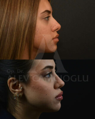
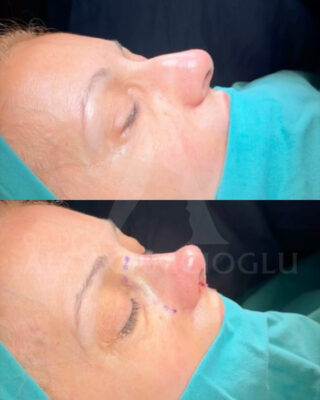
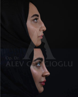
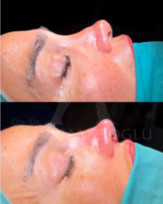
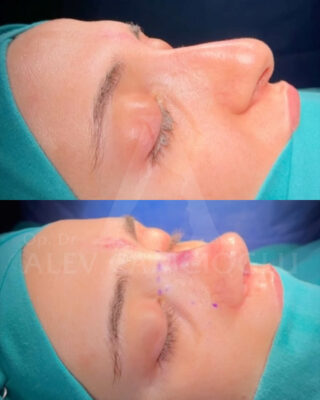
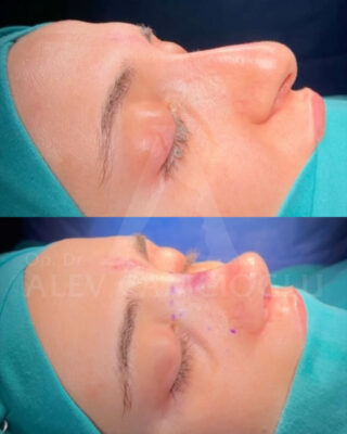
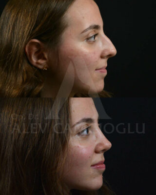
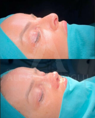
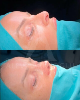
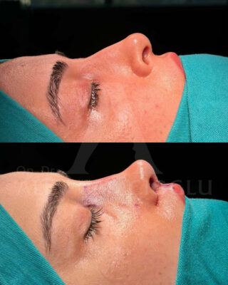
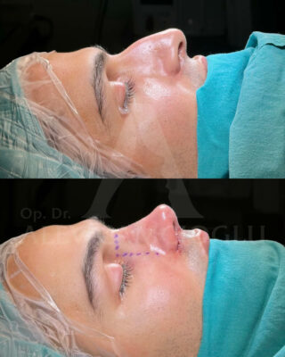
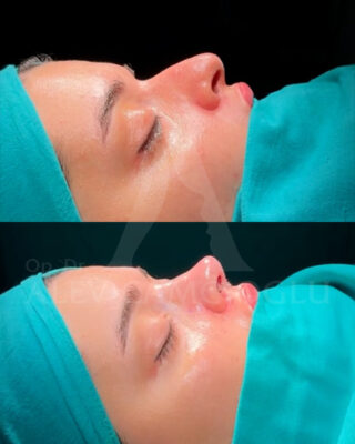
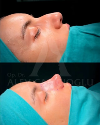
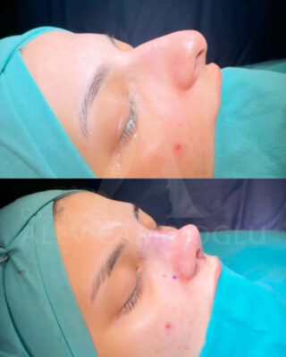
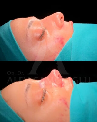
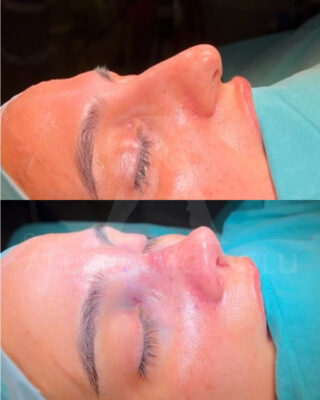
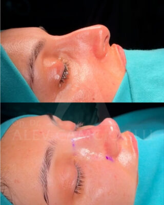
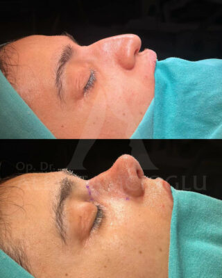
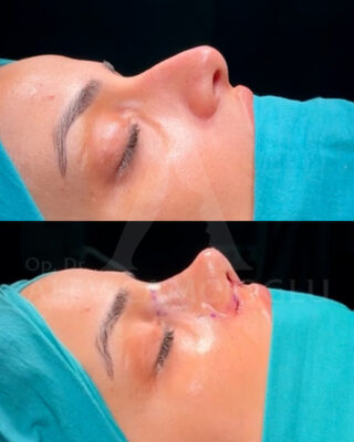
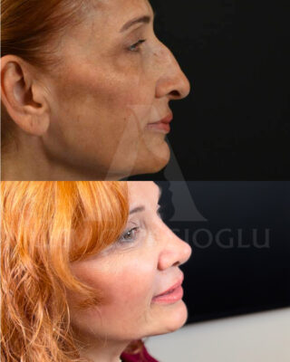
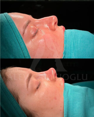
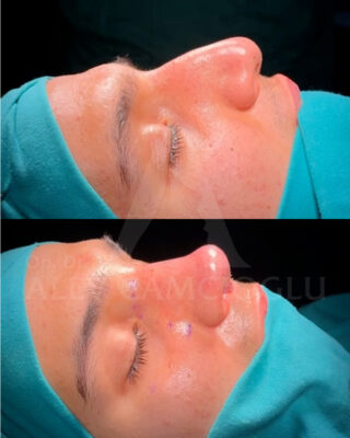
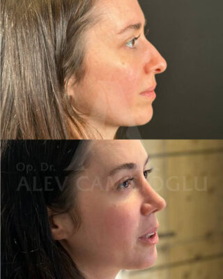
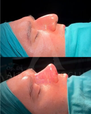
Functional Rhinoplasty Before & After Photos in Turkey
Primary Goals of Turbinate Reduction Surgery
Turbinate reduction surgery is performed to ensure comfortable breathing in individuals suffering from chronic nasal congestion. This surgical procedure is especially important for individuals experiencing long-term nasal congestion and not responding to other treatments. Turbinate enlargement is often triggered by factors such as allergies or infections, making breathing difficult and reducing the quality of life.
This surgical intervention opens the airway, allowing air to flow more freely through the nose. At the same time, by improving airflow, it enables the turbinates to function more effectively in air purification, heating, and humidification. Turbinate reduction surgery is typically applied for the following conditions:
- Chronic allergies
- Long-term nasal congestion
- Chronic sinusitis
The success rate of the surgery is quite high, and patients are generally satisfied with the results. However, since tissue regrowth is possible, repeated interventions may be necessary in some cases. Overall, this surgical procedure significantly reduces breathing problems and enhances daily life quality. Given that each patient’s condition is different, a detailed evaluation before treatment is critically important. This process considers the patient’s medical history, allergy status, and responses to previous treatments.
Techniques for Performing Turbinate Reduction Surgery
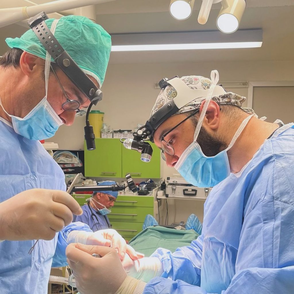
Turbinate reduction surgery is a procedure performed to relieve nasal congestion. During this surgery, doctors reduce the turbinate tissues in the nose to ease patients’ breathing. The procedure is usually performed in hospitals or outpatient treatment centers, mostly under general anesthesia. It can rarely be performed under local anesthesia.
The surgical techniques include:
Cauterization
- A heated probe is placed inside the nose to seal some blood vessels in the turbinates.
- This method reduces blood flow, causing the turbinate tissues to shrink.
Radiofrequency turbinate reduction
- Radiofrequency energy is delivered to your turbinates through a long thin probe.
- This energy causes scar tissue formation in inflamed tissues, shrinking the turbinates.
Coblation
- “Coblation” means “controlled ablation,” and this method also uses heat energy.
- Coblation is performed at lower temperatures to avoid damage to surrounding tissues.
Microdebrider submucosal resection
- The surgeon creates a small opening in the turbinate tissue and removes the tissue through this opening.
- During the procedure, the outer lining is preserved, and the turbinates shrink during the healing process.
Partial resection
- Only a small part of the turbinates is removed during this procedure.
- Partial resection affects both soft and hard tissue.
After turbinate reduction surgery, patients may feel mild discomfort that can last for a few days. It is important to use the medications prescribed by your doctor during the postoperative recovery period. If crusting occurs after surgery, saline sprays or moisturizers can be used for relief.
How is the Diagnostic Process in Functional Nose Aesthetics?
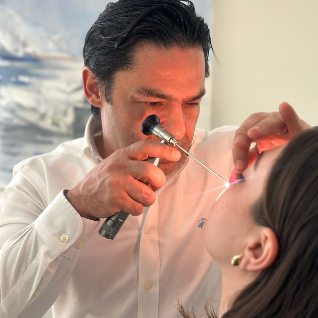
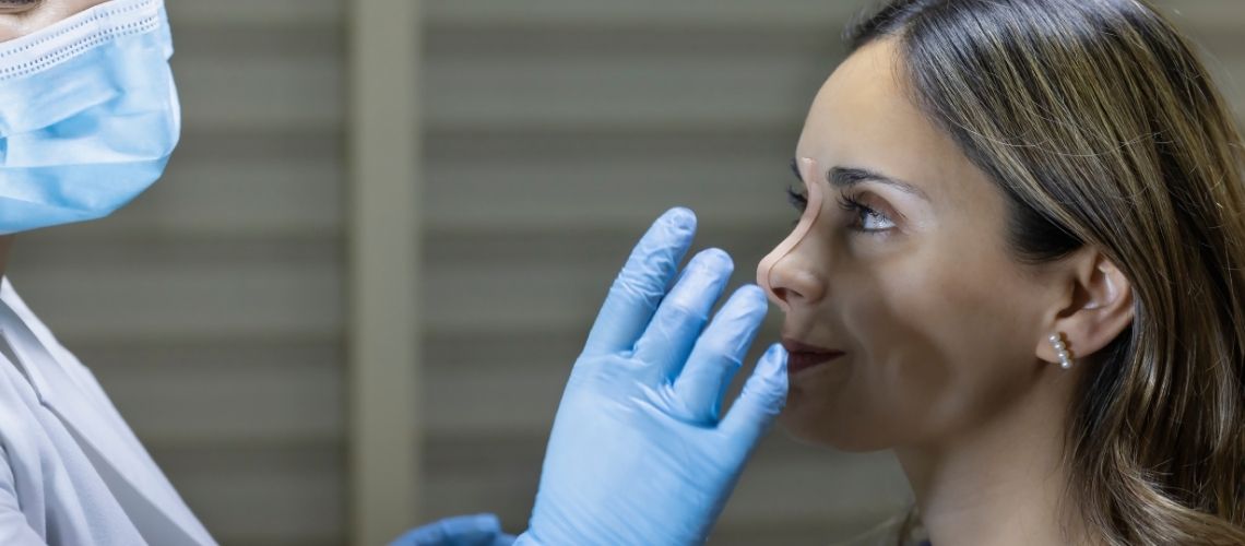
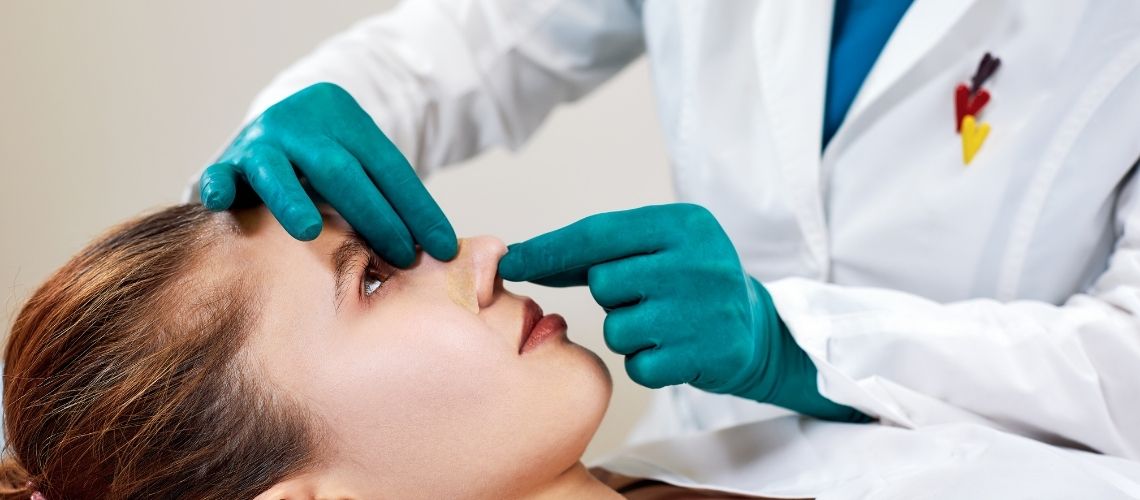
Functional nose aesthetics include a series of evaluations and procedures aimed at eliminating blockages in the nasal airways. This process requires a detailed examination of the patient’s complaints and physical examination findings. The initial evaluation starts with taking the patient’s history, considering past surgeries or traumas that could affect nasal airflow.
During the physical examination, doctors visually and tactilely examine the nasal structure. At this stage, internal and external nasal structural deformities are identified. Some of the techniques used during the examination include:
- Minimal speculum use: A minimal speculum is placed to evaluate the nasal lumen, observing the valves with minimal disturbance to the lateral walls.
- Support with a cotton tip: Intranasal points are supported with cotton-tipped applicators to identify areas that most improve airflow.
During the physical examination, the doctor aims to determine:
- The impact of nasal valve dysfunction on airway obstruction
- Which anatomical structures are insufficient or deformed
- Whether the obstruction is due to static narrowing or dynamic collapse
Examination of the nasal valves is performed both at rest and during normal nasal breathing. This helps differentiate the condition of internal and external nasal valves. Functional assessment is performed in the examination room under active inspiration and with the patient in a supine position. During preoperative preparation, the nasal structure and airflow are carefully examined while the patient is asleep or under sedation.
Operative Techniques in Functional Nose Aesthetics
Functional nose aesthetics aim to address various anatomical and functional problems. The process begins with correcting major deformities of the septum and nasal bones. After corrections are made, the valves are reevaluated. Correcting dorsal septum deviation is particularly important in patients with trauma, as it resolves nasal valve obstruction. Septoplasty to correct anterior septal deviations provides both subjective and objective improvement. Each of these techniques plays a critical role in addressing valve obstruction.
Operative techniques include the following procedures:
- Septoplasty: Corrects septal deviation, opening the airway.
- Osteotomy: Corrects displaced nasal bones, improving valve angle.
- Valvuloplasty: Applied to address valve obstructions.
Osteotomies provide proper reduction, addressing internal valve narrowing. This procedure begins with correcting the cephalic edge of the nasal bones and associated upper lateral cartilages. Properly placed grafts and expansion sutures during the operation support the valves and improve their function. These interventions are typically performed using an open approach, as it allows better visualization and more precise grafting.
Techniques for correcting static and dynamic valve obstructions include:
- Spreader grafts and expansion sutures: Repair static narrowing, widening the valve area.
- Support grafts: Used in areas of dynamic collapse, strengthening the nasal structure.
These techniques are tailored to each patient based on the nasal structure and degree of obstruction. To achieve optimal results, these processes are planned based on preoperative physical examination. However, posterior septal deflections are less frequently corrected as they have less impact on nasal obstruction. Failure to correct them can negatively affect surgical outcomes. Each procedure targets specific deficiencies and is not applied randomly. This comprehensive approach aims to provide patients with healthier breathing.
The Role of Spreader Grafts in Functional Nose Aesthetics
In functional nose aesthetics, spreader grafts are fundamental methods used to increase airway openness. Septal cartilage is considered the most suitable material for these grafts. Standard dimensions are 1 to 2 mm in thickness, 3 to 4 mm in width, and 20 mm in length. If septal cartilage is insufficient, conchal cartilage is used as an alternative.
- Septal cartilage: Usually the preferred material.
- Conchal cartilage: Used when the septum is insufficient.
Correcting internal curvatures is achieved by suturing two conchal grafts with their concave surfaces facing each other. Placing these grafts optimizes nasal airflow. Spreader grafts are placed between the upper lateral cartilages and septum, creating submucosal pockets. This procedure is easiest when starting from the caudal region and progressing toward areas where the septum and upper lateral cartilages join more tightly.
Mucosal elevation allows lateralization of the cartilages, increasing the impact on the internal valve. It is important to avoid damaging the nasal mucosa; otherwise, the graft may become exposed, increasing the risk of infection. If a tear occurs in the mucosa, it is repaired with rapidly absorbable gut sutures.
- Submucosal pockets: Created for placing the cartilages.
- Mucosal elevation: Allows better placement of the cartilages.
- Rapidly absorbable sutures: Used for repairing mucosal tears.
The grafts are secured with a series of polydiaxanone (PDS) sutures extending from one upper lateral cartilage to the other and back again. It is important that these sutures do not go inside the nose, as they could narrow the internal valve. Final adjustments may be necessary to ensure the edges of the spreader grafts are not palpable or visible under the skin. These techniques enhance the success rate in functional nose aesthetics, significantly improving patients’ breathing quality.
The Role of Expansion Sutures
Expansion sutures in functional nose aesthetics play a supportive role in the nasal structure. They are particularly critical for widening the angle of the nasal valve. These sutures are applied to the caudal and lateral regions of the upper lateral cartilage. The sutures extend along the nasal dorsum, securing to the opposite upper lateral cartilage. This procedure directly affects the valve angle, allowing the internal valve to widen.
Application Technique:
- A 4-0 PDS horizontal mattress suture is used.
- The suture extends from one side of the upper lateral cartilage to the other.
- The lateral crus is retracted to reach the optimal fixing point.
Functional Effects:
- As the suture tightens, the upper lateral cartilages are pulled laterally.
- This lateral pull allows the nasal dorsum to function as a support point.
- Tightening the sutures widens the internal valve angle.
A cotton swab is used during suture placement to improve access. This elevates the corner of the upper lateral cartilage, making it easier to place the suture. When used in conjunction with spreader grafts, the expansion suture positions the valve in a more suitable position. Intraoperative assessments show that compared to spreader grafts, expansion sutures significantly widen the internal nasal valve angle. This technique ensures patients breathe more comfortably through their noses.
How Are Suspension Sutures Applied?
Suspension sutures in functional nose aesthetics are used to widen the internal valve angle. During this procedure, the internal valve area is targeted with a surgical Cottle maneuver. The process is supported by a transconjunctival approach, providing access to the infraorbital rim. The suture material used is 3-0 polypropylene, applied in two separate passes through the exposed orbital rim from the endonasal mucosa:
- The first pass is made just above the collapse area of the valve.
- The second pass is approximately 5 mm below the first pass.
The sutures are secured to the orbital rim using three different methods:
- A curved French-eye needle can be used to pass through the periosteum of the maxilla.
- A 1.3 × 6 mm fixation screw can be used to secure the suture.
- A hole is drilled in the orbital rim, and the suture is passed through this hole.
As the suture tightens, the valve area is pulled superolaterally, widening and stabilizing the collapse area. A slight overcorrection is desired at this stage. After releasing the conjunctival flap, it is closed. However, the long-term durability of this suture is still uncertain. Failure to release the soft tissues covering the ULC and LLC may cause the suture to pull out from the soft tissues over time, leading to valve collapse again.
How Do Butterfly Grafts Improve Valve Functions?
Butterfly grafts play a crucial role in correcting nasal valve dysfunctions. These grafts facilitate breathing by maintaining the natural contour of the nasal structure. Both endonasal and open approaches can be used for the application. The grafts are placed between the upper lateral cartilage (ULC) and lower lateral cartilage (LLC). This placement aims to widen the valve angle.
Graft placement:
- In the fold area between ULC and LLC
- To widen the valve angle
- The caudal edge is placed at the cephalic border of the lateral crura
During graft fixation, the material used is 5-0 chromic gut sutures. These sutures are important to prevent the graft from moving. While supporting valve functions, butterfly grafts may also cause some aesthetic changes. These changes generally manifest as a widening of the middle third of the nose. Additionally, slight fullness may occur in the supratip region.
Application Areas and Technical Details of Batten Grafts
In functional nose aesthetics, batten grafts are preferred methods for strengthening weakened or deficient lateral crura (LLC). These grafts are obtained from local sources such as septal and conchal cartilage. Additionally, irradiated homograft rib is used in elderly patients, usually yielding positive results.
The grafts are placed in the soft tissue over the piriform aperture, with their sizes and shapes adjusted according to specific criteria. Graft dimensions generally range from 5 x 25 to 2 mm. To minimize cosmetic effects, the medial side of the grafts features thin beveled edges. To maximize structural support, the battens are kept wider laterally and have notched structures to facilitate fixation.
During preoperative evaluation, the collapse site is identified, a critical step for graft placement. The soft tissue pocket is usually created where the ULC and LLC meet. However, in some special cases:
- After Mohs surgery
- Following other nasal resections
- In cases of lateral wall collapse
The placement of the soft tissue pocket may vary, depending on positions determined during open surgical approaches. After a previous reduction rhinoplasty, excessive resection of the lateral crura often leads to lateral wall collapse, requiring cartilage placement in revision rhinoplasty. These grafts are placed in non-anatomical positions as their locations are adjusted according to existing clinical dysfunctions. The alar batten graft is secured to the nasal mucosa with 4-0 chromic gut sutures.
Causes and Treatment Methods of Tip Ptosis
Tip ptosis is a common issue addressed in nasal aesthetic surgeries. This condition can arise from various problems in the nasal tip structure, and treatment methods are shaped according to the nature of these problems. The treatment process is determined by the anatomical condition of the nasal tip and generally involves the following three situations:
- Structural insufficiencies in the cartilage or ligament structure.
- Excessive soft tissue volume in the nasal tip.
- Vestibular collapse due to lack of forward nasal projection.
In cases of structural insufficiencies, a tip lift suture is used to restore cartilage support. This suture extends from the lower lateral cartilage domes to the periosteum of the nasal bones. The material used in this process is usually 4-0 PDS.
In cases of excessive soft tissue volume, excess skin in the nasal tip is excised to form an aesthetically appropriate scar. This procedure is performed carefully along the boundaries of the nasal subunits. Conditions such as rhinophyma are other causes of soft tissue ptosis, requiring excision in this scenario.
Severe saddle nose deformities require graft use to restore dorsal support. This graft is fixed to the nasal bones and sutured between the lower lateral cartilage domes to improve aesthetics and function. Rib grafts are often preferred in this procedure.
Additionally, other procedures used in the treatment of tip ptosis include:
- Columellar supports
- Lateral battens
- Wedge excisions of the caudal septum
The lateral crural overlay technique developed by Kridel and Konior can correct tip ptosis by shortening the lateral crura and increasing tip rotation. This method is particularly effective in cases where structural and soft tissue laxity associated with aging combine.
How is the Treatment of Paradoxical Lower Lateral Crura Performed?
The paradoxical lower lateral crura issue refers to a condition where the lower lateral cartilages have an abnormal reverse curvature. This condition can adversely affect nasal function and aesthetics. Patients may seek relief from this problem through lateral crural turnover surgery. During the procedure, the lower lateral cartilages are carefully exposed through an open approach.
- Separation from the intranasal mucosa is performed.
- The affected section of the paradoxically curved lateral crus is cut.
- The inverted cartilage is placed in the correct anatomical position.
- It is sutured in place with 5-0 PDS suture.
This technique maintains support from the lateral strut and piriform aperture. At the same time, the integrity of the vestibular mucosa is preserved. Post-procedure, tip grafts are appropriately placed to enhance nasal tip stability.
How Much Does a Functional Rhinoplasty in Turkey in 2025 ?
The cost of a Functional Rhinoplasty in Istanbul, Turkey, typically ranges between €5,000 and €7,000. The price can vary depending on several factors, such as the surgeon's experience, the complexity of the procedure, the clinic's reputation, and additional services included in the package.
| Average Functional Rhinoplasty Cost (in Euros) in 2025 | |
| United Kingdom | €16,600 – €19,000 |
| United States | €14,200 – €23,400 |
| Canada | €16,000 – €19,000 |
| Australia | €23,800 – €37,800 |
| South Korea | €9,400 – €17,200 |
| Ireland | €13,000 – €15,000 |
| Germany | €11,000 – €13,000 |
| Netherlands | €11,000 – €13,000 |
| France | €11,000 – €13,000 |
| Turkey | €5,000– €7,000 |
One of the Best Clinic Team for Functional Rhinoplasty in Turkey
Dr. Alev Camcıoğlu is considered to be one of the best Functional Rhinoplasty surgeon in Turkey. You can check Functional Rhinoplasty reviews here.

Op. Dr. Alev Camcıoğlu
Rhinoplasty Surgeon

Aylin Uyuşmak
Clinical Coordinator

Serra Sevgili
Clinical Assistant

Sema Yapıcı
Operating Room Nurse


